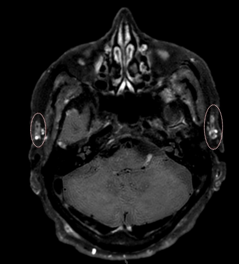
T1-Weighted 3D-Black-Blood-Imaging in Giant-Cell Arteriitis Temporalis and Extracranial Arteritis: A Case Report
Clinical Medicine And Health Research Journal,
Vol. 3 No. 4 (2023),
16 August 2023
,
Page 499-504
https://doi.org/10.18535/cmhrj.v3i4.215
Abstract
Giant-cell arteritis (GCA) is a common vascular inflammatory disorder that often presents with clinical symptoms necessitating prompt diagnosis. Delay in diagnosis can lead to severe patient impairment. This case report highlights the utility of contrast-enhanced T1-weighted 3D-Black Blood (BB) imaging in the diagnostic work-up of GCA, along with its histological correlations. We present the case of an 81-year-old male patient with clinical symptoms suggestive of bilateral temporal arteritis. The patient underwent cranial magnetic resonance imaging (MRI) with contrast-enhanced T1-weighted 3D-BB sequence, which revealed wall-enhancement with perivascular "stranding" of both temporal arteries and their branches, luminal narrowing, and pathological enhancement of the vertebral and basilar arteries. Histological analysis after biopsy confirmed a diagnosis of temporal arteritis.
This case report emphasizes the valuable role of contrast-enhanced T1-weighted 3D-BB imaging in the diagnosis of GCA, providing higher resolution and flow signal suppression. The observed "perivascular stranding" and histological findings contribute to our understanding of the disease. Radiologists should consider incorporating this imaging sequence into their diagnostic protocols when GCA is suspected.

How to Cite
Download Citation
References
- Article Viewed: 0 Total Download


